.jpg)
Researchers in the United States have described antigenic “supersite” within the novel acute coronavirus syndrome 2 (SARS-CoV-2) that may have a significant impact on vaccine design to protect against coronavirus infection. 2019 (COVID-19).
The site was identified on the N-terminal domain (NTD) of the viral spike protein – the main structure used by the virus to bind and capture host cells.
The team – from Columbia University and the National Institute of Infectious Diseases and Infectious Diseases in Maryland – determined cryogenic electron microscopy (cryo-EM) structures for seven neutral antibodies under the direction of NTD.
All seven antibodies were found that targeted one common site at the periphery of the spike protein, thus defining NTD-antigenic supersite.
Lawrence Shapiro and his colleagues say that it is possible that all the strong neutral antibodies that target the NTD spike will focus on this same superstition.
The decisions for vaccine design have important implications, which may lead to the introduction of NTD in forms.
A pre-printed version of the research paper can be found on the bioRxiv * server while the article is under peer review.
Identifies viral sites that are open to nesting
Since the first cases of SARS-CoV-2 were identified in Wuhan, China, at the end of last year (2020), unparalleled efforts have been made worldwide to prophylactic and therapeutic approaches. developed to protect against COVID-19.
One such approach is to identify and study neutral SARS-CoV-2 antibodies to establish sites within the virus that are vulnerable to neutering.
The main target for neutralizing antibodies is the viral spike protein. This structure binds to the angiotensin-converting enzyme receptor 2 (ACE2) on host cells and mediates the virus’s binding to the cell membrane.
The spike protein has two subunits. Subunit 1 (S1) includes the receptor binding domain (RBD), the NTD and several other subdomains, while subunit 2 (S2) mediates virus-cell membrane fusion.
Most of the SARS-CoV-2 neutral antibodies identified so far target the RBD. Studies have revealed neutral antibodies that recognize the RBD at several different sites. They have also shown classes of RBD-led antibodies that appear to be highly elevated, both in human and animal models.
“Although RBD-guided antibodies have been extensively studied, little is known about NTD-directed antibodies,” said Shapiro and colleagues.
However, one study performing EM analyzes of antibodies from convalescent donors identified several NTD-neutralizing antibodies with potency competing with those of the best RBD-guided neutralizing antibodies, add the team.

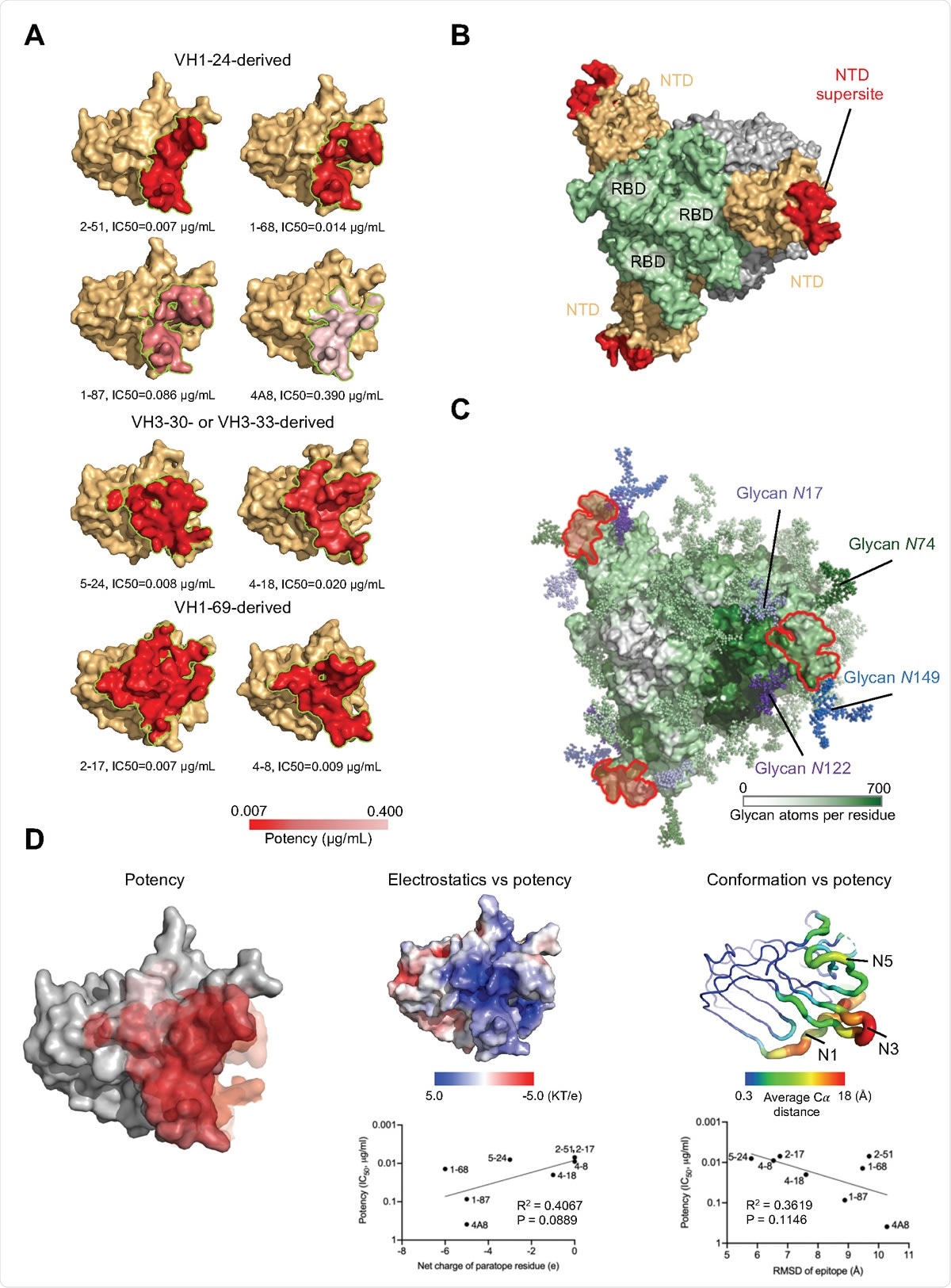
Structured plastic antigenic position in NTD distal loop region revealed by comparing antibodies derived from the four multivariate classes. (A) Epitopes of NTD-targeted antibodies stained with potency. (B) Vulnerability situation on NTD. (C) Glycan coating on the spike. The NTD supersite is encapsulated with glycans at N17, N74, N122 and N149. (D) NTD structural properties and antibody strength. The epitope surface of various antibodies was coated on NTD with red scales representing strength (left). A directional correlation was identified between antibody potency and epitope electrostatics (middle) and conformal change (right).
What did the current study cover?
Shapiro and colleagues have described cryo-EM and crystalline structures for seven neutron-neutral antibodies in complex with either the spike protein or isolated NTD.
The team analyzed the genetic basis of identification for the antibodies and classified them based on their genetics and identification methods. Several classes of antibodies have been tested, and at least one of them has been observed in several convalescent donors.
Surprisingly, examination of the structures showed that all seven antibodies were targeted at a single site on the NTD. This site was a large glycan-free surface located at the periphery of the spike protein and overlooked from the viral membrane, explaining NTD-antigenic superstructure.
“While RBD reveals many unrelated sites of antibody vulnerability, it appears that NTDs are only one vulnerable site to neutralize,” the team wrote.
Shapiro and colleagues say this may be due to the high glycan density on the NTD, with very little glycan-free surface that the immune system recognizes.
“We suggest that all NTD-administered SARS-CoV-2 neutral antibodies could target this site,” they say.
What is the impact of the study?
The researchers say the study has identified a site of vulnerability for NTDs that are more likely to be targeted by neutralizing antibodies and that there are many ways to incorporate it into vaccine design. . Formulas may contain NTD in addition to the RBD and the supersite NTD may be exhibited on nanoparticle immunogens, e.g.

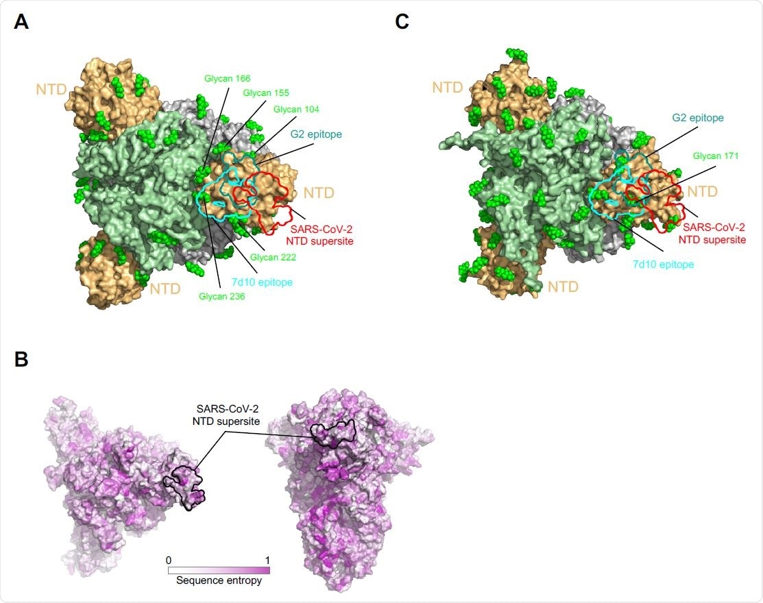
NTD conditioning on MERS betacoronaviruses. (A) Epitopes of MERS NTD antibodies target a site closer to the trimer axis. Epitopian borders of antibody G2 and 7d10 are teal and cyan in color, respectively. SARS-CoV-2 NTD superstar is shown as a red finish line. Glycans are shown as green fields. (B) Spike series entropy between betacoronaviruses. (C) NTD of HKU1 spike is highly glycosylated.
In terms of therapeutic modalities, this superstition is isolated from the sites targeted by RBD-directed antibodies, thus allowing for joint neutralization.
“With all, or the vast majority of NTD-neutralizing antibodies, targeting a single site, however, it appears that there may be little convenience in using a combination of neutralizing antibodies. led by NTD, ”note the researchers.
* Important message
bioRxiv publish preliminary scientific reports that are not peer-reviewed and, therefore, should not be seen as final, guiding health-related clinical practice / behavior, or be treated as information established.