The 2019 pandemic of coronavirus infection (COVID-19) has caused significant damage to human life and well-being. The current problems are unlikely to end if population protectionism is not achieved. The causative agent of COVID-19, the severe respiratory coronavirus-2 syndrome (SARS-CoV-2), has spread to all regions of the world, with more than 106 million registered infections and more than 2.3 million injuries to date. seo.
A team of researchers recently studied SARS-CoV-2 transcription and site-specific dynamics of furin S gene expression in primary human airway epithelia. The team is on their findings about the bioRxiv* preprint server.
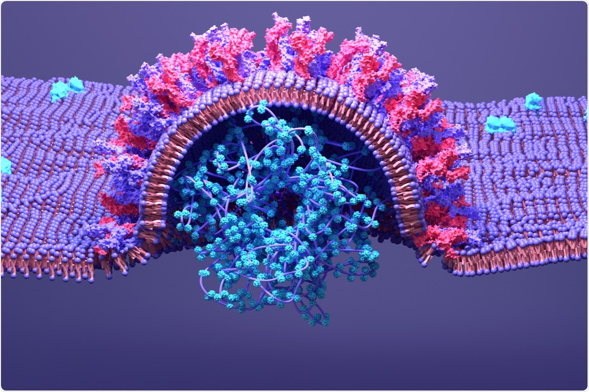
Spike SARS-CoV-2
This virus has a large 30 kb genome, encoding its structural and non-structural proteins, as well as several accessory proteins. The spike proteins of the various coronaviruses share a homology of 77%.
SARS-CoV-2 spike proteins have two subunits, the S1 and S2. S1 mediates an angiotensin-converting enzyme 2 (ACE2) cell spike-host receptor, while S2 mediates viral entry into the host cell through membrane fusion.
The spike protein is the immune antigen in human infection, as well as playing a key role in the type of host cell that the virus targets, the pathogenity of the virus, and its ease of transmission. In nature, it occurs as a homotrimer on the surface of the viral piece.
Importantly, the SARS-CoV-2 spike differs from the SARS-like coronaviruses that are closely related to the codification of the polybasic fur (FCS) site at the S1 / S2 interface. This is made up of four amino acids, PRRA.
The FCS gene itself is composed of the RRAR ↓ S sequence. Its presence in this virus, but not in other betacoronaviruses, indicates that it is essential for the virus to reactivate. increase in the host area, the range of cell tropism, transmissibility, and pathogenity.
A quick look at deleting FCS
When grown in Vero cells, derived from African green monkey cells, SARS-CoV-2 rapidly underwent S1 / S2 interface mutations that counteracted the loss of the FCS with the proximal amino acids and distal in the generation filters. These created a stable stable population with repeated movement.
The spike gene is the most variable among all SARS-CoV-2 species, and the FCS region at the S1 / S2 interface is particularly susceptible to mutations. A combination of deletions and mutations has been identified in this region in Vero cells by the virus.
In particular, a single mutant with the elimination of a pair of 30 coins was able to more efficiently reproduce within Vero cells, becoming a mainstream on a corridor. Another 21-coin pair was also eliminated after two or three passes. These were not detected in the initially prescribed clinical isolation, however. This indicates that these were obtained during Vero cell passages.
Clinical isolations show that there is a lot of erasure around the FCS. The current study aimed to understand how these arose during the virus’ growth in human airways.
Study details
The researchers used RNA sequencing techniques to study virus transcription in human airway epithelium (HAE) cells, implanted to mimic the natural susceptibility of the human airways. This was achieved by cultivating them at an air-liquid interface (HAE-ALI).
This culture approach produced a more similar transcription pattern in the normal human airway epithelium in COVID-19 patients. In both HAE-ALI and Vero cell cultures called SARS-CoV-2, nucleocapsid (N) transcripts, in the form of subgenomic RNA (sgRNA), were abundant, with lower levels of spike transcripts (S ).
They found that large differences largely involved elimination at the S1 / S2 interface. The high rate of FCS eradication contributed to a much lower percentage of sgRNAs compared to all canonical sgRNAs, at only 50%, in infectious HAE-ALI cells.
Two deletions in the FCS were found at rates of significant increase in two cultures. One is the elimination of 12 amino acids (mut-del1), 678TNSPRRAR ↓ SVAS689, including the FCS. The second is the elimination of 5 amino acids (mut-del2), 675QTQTN679, just upstream of the FCS.
Special donor selection
They monitored selective stress specific to donors working within these HAE-ALI cells to eliminate or select such. This was confirmed by the presence of mut-del2 in 42% of infectious cells at 21 days post-infection, and mut-del1 in 54% at 13 days post-infection, in two of seven different cell cultures based on cells obtained from different donors.
Mut-del1 may be more clinically important than mut-del1, having been detected in three of 68 samples in an earlier study.
On the other hand, FCS eradication was eliminated in five other cultures, with less than 1% eradication detected by day 13 after infection. Thus, HAE-ALI cultures from different donors continued to select different FCS eliminators.
Deletion of FCS and viral entry
Mut-del2 will not result in the loss of the FCS, but it does prevent the use of furin media due to the upstream simulation. As a result, spike processing was adversely affected. As a result of both deletions it was not possible for you to use either furin or TMPRSS2 to perform the fusion of the virus with the plasma membrane, thus preventing viral entry into the cell.
The range of cell tropism was also limited by the elimination of FCS. Cells expressing the presence of TMPRSS2 did not show the elimination of FCS, supporting the role of the latter in eliminating the ability of the first to mediate virus-cell fusion.
However, FCS mutations accumulated among new viruses in two cell cultures. These were the only airway stem cells that showed increased levels of mutants, indicating that they are susceptible to infections by mutants depleted by FCS. This may be because, with a high lack of TMPRSS2 sensitivity, they allowed infection with other pathways that involved cathepsin-dependent endosomal entry.
Also, the results from mut-del2-related reductions in viral entry and infectivity in Vero cells may indicate that FCS is a factor of violence. In this case, it should reduce lung disease in animals. Therefore, further study is indicated to understand the frequency of such eradication in human disease, how such mutants are transmitted between humans, and the degree of pathogenity.
. At 4 days after infection, a piece of the insertion membrane was immersed in 4% paraformaldehyde in PBS at 4 ° C overnight. and subject to direct immunofluorescence analysis. The organs were stained with anti-SARS-CoV-2 N (NP) protein. The Leica TCS SPE confica microscope was photographed under 40 ×, controlled by Leica Application Suite X software. The nuclei were fired by DAPI (4 ′ =, 6– diamidino-2-phenylindole). There is a 20 µM scale bar.")
Immunofluorescence analysis of SARS-CoV-2 infection in HAE cells. HAE-ALIB2-20 cultures were infected with SARS-CoV-2 at an MOI of 0.2 or 2 pfu / cell, as indicated, mock-disease (Mock). At 4 days after infection, a piece of the insertion membrane was immersed in 4% paraformaldehyde in PBS at 4 ° C overnight. and subject to direct immunofluorescence analysis. The organs were stained with anti-SARS-CoV-2 N (NP) protein. The Leica TCS SPE confica microscope was photographed under 40 ×, controlled by Leica Application Suite X software. The nuclei were fired by DAPI (4 ′ =, 6– diamidino-2-phenylindole). There is a 20 µM scale bar.
What is the impact?
These results indicate that there is a strong selective pressure on the virus within human airway cells, for variable mutations in the spike protein. Vigilance to identify spike mutations is essential to identify escape mutations, as well as those that affect or exacerbate viral infection or transmission, or violence. The current study succeeded in the HAE-ALI polarization model to study spike mutants in several cases.
The researchers found that a selective force in viral grains apically released from polarized human airway cells resulted in stable retention of the FCS and total spike stability in most cases, except two cultures that were separated from two different individuals. In both cases, high levels of FCS deletions followed.
Our study includes evidence for the role of specific human airway epithelia properties in FCS area dynamics when human airways are dependent, donor-dependent. ”
* Important message
bioRxiv publishes preliminary scientific reports that are not peer-reviewed and, therefore, should not be seen as final, guiding health-related clinical / behavioral practice, or treated as fixed information.