“For too long, continuous imaging of brain tumors has been done as a speculative idea rather than a science-based schedule,” said lead author Dr. Tom Booth, PhD, consultant neuroradiologist at King’s College Hospital NHS Foundation Trust, London. AuntMinnieEurope.com. “A large group of experts was encouraged to make a position statement to explain the status quo and use it as a springboard for good study, allowing for a science-based timeline.”
The document was published in the journal Open Access Frontiers in Oncology. It came from a meeting held in London in April 2019 attended by representatives of UK stakeholders to test an approach to interim imaging of adult brain tumors. Representatives of charities, neuro-oncologists, neurosurgeons, neuroradiologists, neuropsychologists, testers, health economists, data scientists, and the imaging industry were all represented. It was held in conjunction with the Brain Tumor group workshop at the National Cancer Research Institute (NCRI).

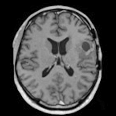
The figure shows long-term images after treatment. The timing of scans is a practical solution to assess treatment response, but the approach is not evidence-based and the images do not always give a definitive correlation as to whether the tumor is coping. treatment. We don’t know for sure whether we image too often, too little, or the right amount in terms of morbidity (including quality of life), mortality, and cost-effectiveness, Dr. Tom said. Booth, PhD. With the permission of Dr. Tom Booth, PhD.
Clinicians tend to use brain scans at preset times to determine if a brain tumor patient is responding to treatment, but the frequency of scans can range from every few weeks to every few months. . Different countries and hospitals use different approaches, but determining the best science-based approach is a real challenge.
“I felt this was really needed and Dr Robin Grant, a pathologist with an interest in adult brain tumors, suggested that the best form would be through Incubator Day,” Booth explained. on the status quo and look at how to gather evidence in the future. “
Gaps in evidence
The situational narrative describes why brain imaging is a largely neglected area, and the authors clarify the holes in the evidence and explore ways to gain the necessary knowledge.
The study of early adult brain tumors has always focused on therapeutic clinical trials, and images as an “add-on” to the clinical trials were seen as a practical solution to the problem of trying to assess whether the treatment working or not, according to Booth.
“Like much of the radiation (and medicine), the rationale for the management of such patients based on imaging has never been thoroughly studied: This is often because it is challenging to do so,” he said. e. “An additional reason not to question the status quo is that it has become so rigid in routine practice and routine clinical trials that it would be a major challenge to redesign interim images if the evidence shows that the conventional imaging practices are not appropriately causal. “
More collaborative efforts are now needed to make progress, Booth continued.
“This is because the complexity of treatment and the scarcity of brain tumors require that so much data and information be brought together,” he said. “There are a number of collaborations looking at different aspects of the patient pathway – usually from a therapeutic or surgical point of view. When it comes to imaging, radiologists usually deal with one area at a time. “
An important example here is the study entitled “Development of Glioblastoma Treatment: Distinguishing Progress from Pseudoprogression,” where more than 10 facilities in the UK collect imaging and clinical imaging data. The organizers hope that this government-backed machine learning collaborative experience will be used to address the larger issue of making full use of the entire imaging trail.
In addition, there are some potentially promising UK databases on the horizon, including BRIAN (prospect data) and MATRIX (prospective data).
Global relevance
The project started as a UK-focused position statement and involved all UK brain charities, so there was no overseas involvement, but Booth is sure the campaign will be interesting. and relevant to overseas colleagues.
“It is clear that the type of health care system is likely to influence the use of interim images through, for example, the type of national reimbursement system,” he said. global evidence – as the references prove – and it is written in a way that will be used around the world with reference to different health care systems. “
The recommendations for future study are not didactic and are common to a range of health care systems, he said.
Overall, Booth hopes the global image community will make use of the statement. The recommendations for future study are valuable to everyone, and are common to a range of health care systems. The wide range of proposed future studies, and how they might build the required body of evidence, is particularly relevant to all researchers in this field, according to Booth. .
“Clearly pathways need to be increased so that we can scan the right patient at the right time with a number of clinical benefits downstream: this is true before, during, and after COVID,” he said. -19 has clearly led radiologists and other clinicians to question the evidence behind their activity even more because the risk benefit of being present for a scan has changed now. “
Copyright © 2021 AuntMinnieEurope.com