Nothing fills a parent with relief like the sound of their child’s cry. That first air gulp describes such a critical moment in our lives, yet it happens to be one of the least understood of any human behavior.
Most of what we know about physical activity within the lungs during the first breath comes either from preclinical studies – often involving animals – or through being taking an aggressive eye that is in danger of disrupting natural lung functions.
Now, using a delicate sensor belt wrapped around newborn full-time chests after delivery, a team of researchers from Australia, Germany, Switzerland, and Canada have been on the record changes in lung volume that occur in the first minutes outside of infancy. inside.
“Healthy term babies use highly complex methods to adjust to airway breathing at birth,” says clinical neurologist David Tingay of the Murdoch Children’s Research Institute (MCRI) in Australia.
“There’s a reason parents, midwives, and obstetricians are happy to hear those first life-affirming calls when a baby is born.”
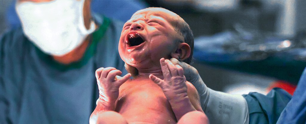
The machine created images of baby coffins, which combined video and audio data with detailed descriptions of more than 1,400 breaths taken by 17 tiny people all delivered through a selected cesarean section.
The results provided a statistical overview of the process of lung ventilation in neonates – a physiological activity in the preparation and insertion of lungs to allow them to take the place of the placenta for exchange gases.
Put simply, while the babies are sucked into the uterus, they are busy using their lungs: Although they are full of fluid that is peeled by the lining of the lungs, they still movement in temporary breath to put the brain and muscles to good use. .
This training regime falls just before birth. The intense pressure that comes with being pushed through the vaginal canal removes some of the fluid, while at the same time providing a generous dose of adrenaline that urges the lungs to absorb as much of that liquid as they can.
That still leaves a good chunk of debris going around, especially if the delivery came by caesarean. Much of it is pushed back through the figs with the first air ash, with a soapy substance called a surfactant helping the tiny airbags peel off the wetness cover and expand it as wide as possible.
No wonder the first few times we swim around outside come with a gurgling cry.
Data from this new study will help dispel the myth. This weeping creates a distinct pattern of gas flow that helps maintain the residual capacity of action; the air we hold in our lungs after we breathe.
There was no need to balance this breath between the two lungs either, tending to favor the right side more than the left.
Of all recorded breaths, crying was mostly present in the first minute after birth when babies were shrinking their lungs; 6 minutes later, more than half of the baby’s breath had moved into a steady stream called tidal breath.
Having signatures of breathing patterns in healthy infants delivered by caesarean provides a basis for studying how lung ventilation differs in other cases, such as in preterm infants.
About 1 in 10 newborns, and almost all babies born before 37 weeks, need help to catch that first important breath.
For those who find this type of research very interesting, MCRI has produced a detailed, yet easily accessible video clip, the study explains, which you can watch below.
“The development of delivery room interventions must first be understood in the processes that explain the success and failure of breathing at birth,” says Tingay.
In emergencies this may require traumatic interventions such as intubation, which can – although save lives – damage airways and leave babies at risk of cigarette damage. airway, infection and respiratory diseases.
Knowing when to take action, and when not, can help put new babies at unnecessary risk.
The equipment used in the study is about as non-invasive as it gets, so it could be applied in most births, caesareans and vaginal implants, and not there is little danger to the new baby.
“Not only does this new technology allow us to see deep into the lungs but it is also our only way of making continuous images of the lungs without having to ‘using radiation or blocking life-saving care,’ “Tingay said.
“This study has shown that babies’ lungs are much more complex than traditional screening methods have previously suggested.”
This research was published in the American Journal of Respiratory and Critical Care Medicine.