One cannot look inside someone else’s head. This makes it difficult to understand what is happening in the brains of people with dementia. A new approach offers insight.
A team of researchers from Ruhr-Universität Bochum (RUB) and Vrije Universteit Amsterdam (VU) have tested the developmental stages of Aβ fibrils and thus been able to understand the development of plaques. The researchers report in the academic journal Communication Acta Neuropathologica on 11 December 2020.
Coming together
The team from the Center for Protein Diagnostics (Prodi) at RUB is working with Vrije Universiteit Medical Center (VUmc) to bring together expertise in protein research and depression. Medical specialists like Baayla Boon from VUmc attend to patients with depression in the clinic and examine post mortem brain tension, which is diligently collected by the Dutch Brain Bank (NBB). Thin razor slices are cut from the brain for examination. “Such chips allow us to look deep within the determinants of the brain,” says Baaylas director, Annemieke Professor Rozemuller, Head of Neuropathology at VUmc.
The groups exchange samples and conclusions at regular meetings. In Bochum, protein researchers led by Professor Klaus Gerwert, Head of Biophysics and Prodi’s founding Director at RUB, study brain tension. The tablets here are illuminated by infrared light in special microscopes. “This will allow us to test the packaging of biomarker proteins in the strain,” says Dominik Röhr from Prodi.
New timeline for records
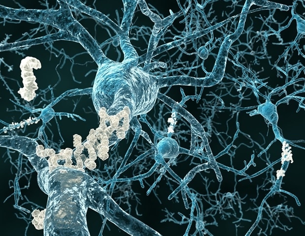
The formation of Aβ fibrils is notorious for laboratory experiments. First, Aβ folds like a sheet of paper to form so-called β-sheets. These gather together in small groups, called oligomers. Over time, the β-sheets come together as a deck of cards to form fibrils. The Bochum-based researchers led by Klaus Gerwert use this process as an internal clock for records.
There are many oligomers in newly developed records. Over the course of plaque development, fibrils are formed continuously. The researchers therefore concluded that records go through different stages during their development.
It has not been possible to monitor pest development directly to date. Combining techniques from medicine and physics, new opportunities are now opening up. ”
Professor Klaus Gerwert, Head of Biophysics and Founding Director of Prodi at RUB
On the left is a fully developed tablet of Aβ with brown color. On the right, the structure of Aβ confirmed by an infrared microscope is shown. In the heart are fibrils (red), mostly surrounded by oligomers (blue).
Aβ is the focus in the fight against Alzheimer’s disease. A key approach in finding a cure is to spread the plaques in the brains of the patients. “Early diagnosis of Alzheimer’s disease is crucial to prevent Aβ from causing unstable brain damage,” says Dominik Röhr. Promising drugs are currently under licensing testing. These include the antibody aducanumab, which can break down plaques in the brain.
The new findings from an infrared microscope indicate that the development of pests could be stopped at an early stage by inhibiting the formation of oligomers. These are thought to be particularly harmful to the brain. The toxic effect of Aβ could therefore be reduced by appropriate drugs.
Source:
Magazine Reference:
Röhr, D., et al. (2020) Unlabeled animated images of different types of Aβ plaque in Alzheimer’s disease show sequential events in plaque development. Communication Acta Neuropathologica. doi.org/10.1186/s40478-020-01091-5.