Capturing details of brain cells at the nanometer scale, researchers have found evidence that the neurons of people with schizophrenia may have distinct differences in thickness and curvature, and this may even account for some of their symptoms.
The result comes from a study of only a handful of donors, and is far from showing how different zero-cell structures could explain the neurological condition.
But as our understanding of these unusual features grows, this could lead to better treatments, helping to deliver a better quality of life for tens of millions across the world. world.
The study, led by researchers from Tokai University in Japan, made use of two different X-ray microscope technologies, one at the SPring-8 light source facility in Japan, and the other at Advanced Photon Source US Department of Energy (APS).
Both of these accelerate particles on curved paths in what is called a synchrotron, causing them to emit short waves of electromagnetic radiation in the X-ray part of the spectrum.
Using X-rays as a source of radiation for fine images of tiny objects – such as neurons – can be a bit of a two-edged sword.
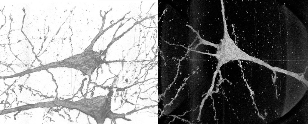
On the one hand, their tight waves are just the thing for capturing every buckle and weave of a cell membrane. The APS is capable of dissolving down as far as 10 nanometers, a scale that brings it very close to revealing the texture of individual protein channels that hit a cell.
Viewed from sufficient angles, it is possible to reconstruct neurons as high-definition, three-dimensional domains.
Unfortunately, as tiny as neurons, they are also very long. Finding every lump of their surface is a daunting task when you need to travel your way across the entire millimeters of the body.
“The sample has to move through the X-ray beam to find the neurons through the sample,” says Vincent De Andrade, a physicist in Argonne’s Department of X-ray Science.
“The field of view of our X-ray microscope is about 50 microns, about the width of a human hair, and you have to follow these neurons over several millimeters.”
Taking samples of material from a selected part of the brain in four deceased people diagnosed with schizophrenia, and four without, the team performed the tedious task of scanning zero cells using both synchrotron devices different.
The images were combined to reconstruct the neurons as digital models, contributing to a larger data set that could be statistically compared and compared in search for different features .
They found that, statistically, the thickness and curvature of cell features extending away from the body of the neuron were significantly different among people with schizophrenia, compared to those without the condition.
These changes could affect how the neurons transmit messages down the length, which may go a little further in explaining the features of the disorder, which are in the worst forms. his includes hallucinations, obstruction of motor control, and convulsions.
Exactly what is behind such movements in cellular geometry, or whether the changes extend all the way to the synaptic ‘toes’ of the neuron, will require even more precision than normal generation synchrotrons can to govern.
That could change when the APS receives a US update of $ 815 million in the next few years that will deliver X-ray long haul 500 times brighter than its current releases.
“Updating APS will allow better sensitivity and resolution for images, making the process of mapping neurons in the brain faster and more accurate,” says De Andrade.
“We needed resolutions better than 10 nanometers to capture synaptic connections, which is the sacred magnitude for extensive mapping of neurons, and those should be achievable with the upgrade.”
Putting the devices behind the development of schizophrenia is a complex process that requires advanced technology and computer technology.
Gradually we come to understand the number of genetic and environmental factors that see the brain change while they are still in the womb, and continue to move as a child grows. to become an adult.
If there are ways that this can be seen and treated early, we could help to limit, if not stop, the worst symptoms that can put people at risk of serious mental illness.
This research was published in Translational psychology.