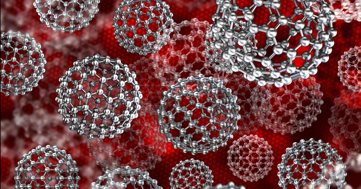
Nanoparticles, which are increasingly used in modern biomedicine, form biomolecular coronas on their surface that interact with the biological environment around them. The biomolecular corona has previously been defined as an undefined and diffuse network of proteins that bind to nanoparticles. Proteins are well studied due to their wide range of cell activity and advanced technologies for analysis (eg tandem mass spectroscopy liquid chromatography).
“Taxpayer money has been heavily invested in cancer nanomedicine research, but that research has failed to reach the clinic,” Morteza Mahmoudi, PhD, assistant professor in the department of radiology at Michigan State University, said in the recitation. “The biological adverse effects of nanoparticles, how the body interacts with nanoparticles, are not understood. And they need to be considered in detail.”
New ways to illuminate the structure, circulation, homogeneity and interactions of biomolecules near the surface of nanoparticles are needed. Mahmoudi’s team developed a mixed-method microscopy method (combining cryo-electron microscopy, electron cryotomography, 3D reconstruction and image processing, and image simulation) that allowed researchers to look at biomolecular coronas more accurately than ever before. The team used carboxylated polystyrene nanoparticles for the analysis.
The specific properties of each nanoparticle play an essential role in the formation of the biomolecular corona, which has been confirmed in the present study. For example, nanoparticles influence the size and cost of the biomolecular corona surface.
The researchers found a random rotation and density of biomolecules adsorbed (retained) on the surface of the nanoparticles. This reinforces the understanding that minute changes in small-molecular-weight drugs can have a significant impact on patients ’biology and efficacy.
“For the first time, we can visualize the 3D structure of the grains coated with biomolecules at the nano level,” Mahmoudi said. “This is a useful approach to obtain helpful and robust data for nanomedicines, to obtain the kind of data that will influence scientists’ decisions about the safety and efficacy of nanoparticles.”
When nanoparticles were exposed to human plasma, they stimulated several reactions, many of which are classified as nonspecific proteins. The researchers explained that other analytical methods for proteomics may lead to the incorporation of these nonspecific proteins, leading to an expected action of the corona that differs from its actual activity in vitro. and in vivo.
A combination of the magnetic levitation and cryo-TEM construction method was used to visualize the bonding of individual biomolecules with the surface of individual nanoparticles at the nanoscale.
The team was also able to obtain 3D images of the proteins and biomolecules within the biomolecular corona and their relationship to the nanoparticles using electron cryotomography. The images showed a random and inconsistent collection of medium-sized records of biomolecules. The authors noted that individual molecules could not be resolved. Biomolecules appeared to be both attached to and at distances from the surface of the nanoparticles.
While much remains to be learned, there are opportunities for the nanoparticles in a number of applications, according to Mahmoudi. For example, instead of trying to treat diseases with nanoscale treatment, he believes that nanoparticles would be well-suited for early detection of disease such as cancers and neurodegenerative diseases.
Do you have a particular perspective on your research related to drug development or biochemical engineering? Contact the editor today to learn more.
—
Copyright © 2021 scienceboard.net