A new noninvasive magnetic resonance spectroscope device could help providers measure how well treatments are working for patients with amyotrophic lateral sclerosis (ALS), a new study has found.
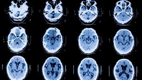
31-phosphorus magnetic resonance spectroscopy is specifically known as a measure of how well the mitochondria are functioning in patients with this motor neuron disease (MND), also known as ALS. Researchers from the Sheffield Institute for Translational Neutralization (SITraN) from the University of Sheffield in the United Kingdom tested how well this imaging method works and published their findings in the journal Jan. 13 de Brain.
“In this study, we found that phosphocreatine levels were lowered in the brain compared to healthy controls and, in this neck, we found that inorganic phosphate was elevated in patients with MND,” said the author of the lead study, Dr. Matilde Sassani, neurodegeneration researcher. at Sheffield. “Both of these findings are consistent with mitochondrial intolerance occurring in those living with MND.”
Since existing research shows that mitochondrial activity is impaired in people with ALS, they may have a method that can effectively and accurately measure how well they are doing. it works in living patients to be critical for treatment efforts, she said.
For this study, the researchers used the imaging method to measure chemicals that are heavily involved in cell energy metabolism. They tested both in patients with ALS and healthy controls by age and sex. Although the procedure is strongly similar to a standard MRI test, the process allows investigators to capture a direct amount of chemicals, giving them the data they need to get a complete picture of a patient’s energy status. Work out MND.
According to colleague Sassani, senior author Dr. Thomas Jenkins, senior clinical lecturer at SITraN, said this new ability to assess mitochondrial function could pave the way for new, more effective MND treatments. It could measure how well medications work on mitochondrial function in this patient group.
“Remedies aimed at saving mitochondrial function in MND are being studied in laboratories around the world,” he said. “This non-invasive tool can demonstrate whether developmental drugs are successfully targeting mitochondria, which is an important step in selecting treatments for clinical trial. ”
Future research is also needed, the team said, to test whether this imaging technique has been applied to groups of patients with other types of neurodegenerative disease.
For more coverage based on the opinions of research and research experts, subscribe to the Diagnostic Imaging e-newsletter here.