Press release
Wednesday, December 30, 2020
Results from a study of 19 deceased patients indicate that brain damage is the result of patient illness.
In an in-depth study of how COVID-19 affects patients’ brains, researchers at the National Institutes of Health routinely saw signs of damage caused by thinning and leaky brain blood vessels in print samples from patients who died shortly after catching the disease. In addition, they showed no signs of SARS-CoV-2 in the print samples, suggesting that the damage was not caused by a direct viral attack on the brain. The findings were published as a publication in the New England Journal of Medicine.
“We found that the brains of patients who become infected with SARS-CoV-2 may be susceptible to microvascular blood vessel damage. Our findings suggest that this may be caused by the body’s inflammatory response to the virus ”said Avindra Nath, MD, clinical director at the NIH National Institute on Neurological Disorders and Stroke (NINDS) and lead author of the study. “We hope these results will help doctors to understand the full range of problems that patients may face so that we can better treat. ”
Although COVID-19 is mainly a respiratory disease, patients often experience brain problems including headache, delirium, cognitive dysfunction, dizziness, fatigue, and loss of sense of smell. The disease could also cause strokes and other neuropathologies.
Several studies have shown that the disease can cause inflammation and damage of blood vessels. In one of these studies, the researchers found evidence of small amounts of SARS-CoV-2 in the brains of some patients. Nevertheless, scientists are still trying to understand how the disease affects the brain.
In this study, the researchers conducted an in-depth study of brain tension samples from 19 patients who had died after experiencing COVID-19 between March and July 2020. Samples from 16 of the patients were provided. by the Office of the Chief Medical Examiner in New York City while the other 3 cases were referred by the pathology department at the University of Iowa College of Medicine, Iowa City. Patients died at a wide range of ages, from 5 to 73 years old. They died within a few hours to two months of reporting symptoms. Many patients had one risk factor, including diabetes, obesity and cardiovascular disease. Eight of the patients were found dead at home or in public settings. Three other patients fell and died suddenly.
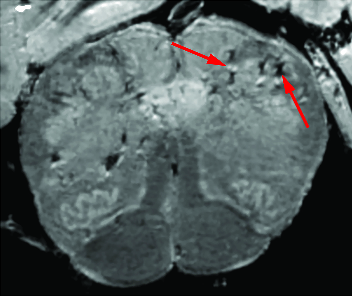
The researchers first used a magnetic resonance imaging (MRI) scanner that is 4 to 10 times more sensitive than most MRI scanners, to examine samples of the olfactory bulbs and brainstems. from all patients. These regions are thought to be highly susceptible to COVID-19. Decorative bulbs control our sense of smell while the brain system controls our breathing and heart rate. The scans showed an abundance of bright spots in both regions, called hyperintensities, which often indicate inflammation, and dark spots, called hypointensities, that represent bleeding.
The researchers then used the scans as a guide to examine the spots more closely under a microscope. They found that blood vessels in the bright spots were thinner than normal and sometimes released blood proteins, such as fibrinogen, into the brain. This was likely to have a protective effect. The spots were surrounded by T cells from the blood and the brain’s own immune cells called microglia. In contrast, the dark spots contained both clotted and leaky blood vessels but there was no immune response.
“It simply came to our notice then. Initially, we expected to see damage caused by a lack of oxygen. Instead, we saw multifaceted areas of damage typically associated with strokes and neuroinflammatory diseases, ”said Dr Nath.
Finally, the researchers found no signs of disease in the brain tight samples although they used several methods to detect genetic material or proteins from SARS-CoV-2.
“So far, our results show that the damage we saw directly to the brain may not have been caused by the SARS-CoV-2 virus,” said Dr. Nath. “In the future, we plan to study how COVID-19 damages the blood vessels of the brain and whether it causes some of the short-term and long-term symptoms. time we see in patients. ”
This study was supported by the NIH Intramural Research Program at the National Institute of Neurological Disorders and Stroke (NS003130) and the NIH grant (NS109284).
NINDS (https://www.ninds.nih.gov) the country’s leading study fund of the brain and nervous system. The mission of NINDS is to seek basic knowledge about the brain and nervous system and to use that knowledge to reduce the burden of neurological disease.
About the National Institute on Aging (NIA): The NIA will lead the U.S. federal government’s effort to monitor and support aging and the health and well-being of the elderly. Learn more about age-related mental change and neurodegenerative diseases through the website of the Alzheimer’s Center and the Center for Dementias Education and Change (ADEAR). For information on a wide range of aging topics, visit the main NIA website and stay connected.
About the National Institutes of Health (NIH):
NIH, the nation’s medical research agency, comprises 27 Institutes and Centers and is part of the U.S. Department of Health and Human Services. NIH is the leading federal agency that conducts and supports basic, clinical, and translational medical examination, and examines the causes, treatments, and cures for both common and rare diseases. For more information about NIH and its programs, visit www.nih.gov.
NIH… Turning traces into health®
Article
Lee MH, Perl DP, Nair G, Li W, Maric D, Murray H, Dodd SJ, Koretsky AP, Watts JA, Cheung V, Masliah E, Horkayne-Szakaly I, Jones R, Stram MN, Moncur J, Hefti M, Folkerth RD, Nath A. Microvascular injury in brain of patients with COVID-19. New England Journal of Medicine, December 30, 2020 DOI: 10.1056 / NEJMc2033369.