Ask anyone, from an NFL quarterback scanning the field for open catchers to an air traffic controller monitoring the position of planes to a parent watching their children run around at the park: It is up to our brain to keep in mind what we see, even as we move our gaze around and even look away for a while. This ability of “visual working memory” feels effortless, but a new MIT study shows that the brain is working hard to maintain. When a main object moves across our field of vision – either because it has moved or as our eyes have done – the brain instantly moves memory by recoding it among neurons in half opposite the brain.
The result, published in Neuron by neuroscientists at the Picower Institute for Learning and Memory, explaining through experiments in animals how we can continuously monitor what is important to us, even though the basic wiring of our visual system requires mapping what we see on our left side on the right side of our brain. and what we see on our right side on the left side of the brain.
“You need to know where things are in the real world, no matter where you look or how you are guided at a particular time,” says study lead author Scott Brincat, a postdoc in laboratory Professor Picower, Professor Earl Miller, lead author. “But the representation of your brain from the outside world changes every time you move your eyes.”
In their experiments, Brincat, Miller, and co-authors found that when an object changes aspects in the field of view, the brain quickly employs a narrative change in brain wave frequency synchronization to decode the memory information from one side of the brain to the other. The movement, which takes place in just milliseconds, employs a new group of neurons in the prefrontal cortex of the opposite brain hemisphere to store the memory. This new ensemble of neurons encodes the object based on its new position, but the brain still recognizes it as the object that used to be in the field of view of the other hemisphere.
It is that ability – to remember that something is the same no matter how it moves around our eyes – that gives us the freedom to control where we look. , Miller says. A quarterback can decide to move his feces from the left side of the field to the right without fear that he will immediately forget that the left catchers are still there, even if the position of the scene changed them to large within, or even out of, its field of view.
“If you didn’t have that, we would be just simple creatures who could only respond to anything that comes right to us in the environment, that’s all,” Miller says. “But because we can keep things in mind, we can control what we do. We don’t have to respond to something now, we can save it for later. “
Moving sides
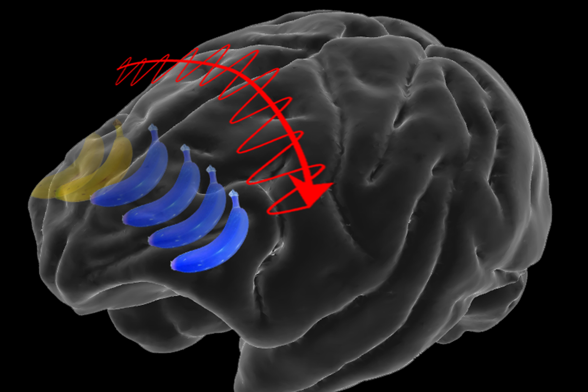
In the laboratory, the researchers measured the activity of hundreds of neurons in the prefrontal cortex of the two brain hemispheres as animals played a game. They had to fix their feces on one side of a screen because an image of an object (eg, a banana) appeared briefly in the center of the screen. So the object appeared on one side or the other of their field of view, and because of a wire across the brain, it was processed in the opposite cortical hemisphere. The animal had to memorize the image, and then state whether a later image was of a different object (eg an apple). In some experiments, however, although the original object was kept as a working memory the animals were cured to change the pitch from one side to the other, effectively changing which side of their field of view. called the memorable statue.
Animals were correct in remembering whether the images given to them matched, but their performance suffered only slightly in cases where they had to move the fetus. Brincat says the mistake suggests he doesn’t have to keep up with the movement as easily to the brain as he seems.
“It feels sad to us, but it seems not,” he said.
To examine their measurements in the brain, the team trained on a computer program called a decoder to identify patterns in the raw data of cloud activity that identified the image memory of the object. As would be expected, that analysis showed that the brain was encoding information about each image in the opposite hemisphere where it was in the field of view. But more surprisingly, it also showed in cases where the animals moved the fetus across the screen, neural activity encoding memory information moved from one hemisphere of the brain to another.
The team also measured the total rhythms of the collecting activity of neurons, or brain waves. They found that the transfer of memory from one hemisphere to the other regularly occurred with a signature change in these rhythms. As the trend unfolded, the synchronization over “low” frequency hemispheres (around 4-10 hertz) and high frequency “beta” waves (~ 17-40 Hz) and alpha “synchronization” / beta ”wave decay (~ 11-17 Hz).
This push and pull rhythm pattern is very similar to one found by Miller’s lab in many studies of how the cortex uses rhythms to transmit information. An increase in the combination of very low and higher frequency rhythms allows sensory information (i.e., representations of what the animal just saw) to be coded or retrieved. An increase in power in the alpha / beta frequency range inhibits that coding, acting as a kind of gateway on sensory information processing.
“This is another type of gates,” Miller says. “This time alpha / beta is experiencing a memory shift between hemispheres. ”
Seeing surprise
Although the rhythm patterns appeared to be consistent with prior studies, the researchers were surprised by another finding of the study: With the same image of material in the same area in the field of view, the prefrontal cortex was employ different neurons if first observed in that location as opposed to moving from the other hemisphere. In other words, animals that saw a banana on the left side of their scene employed a different neural ensemble to represent that memory than they did if the banana was previously seen on the right side. and then move it to that place.
For Miller, the discovery has an interesting effect. Neuroscientists once believed that individual neurons are the basic unit of action in the brain and have recently begun to think that they are ensembles of neurons. The new findings, however, suggest that even the same information could be coded with different, round, ensembles together.
“Ensembles may not even be units of brain function,” Miller thinks. “So what is the unit of action of the brain? This is the place of computing that brain network activity creates. ”
In addition to Brincat and Miller, the paper’s other authors include Jacob Donoghue, Meredith Mahnke, Simon Kornblith, and Mikael Lundqvist.
The research was funded by the National Institute of Mental Health, the Naval Research Office, the JPB Foundation, and the National Institute of General Medical Sciences.