Dr. Ewa Jarocka talks to News-Medical about our senses, and how fingerprints can enhance our consciousness.
What inspired your research on our senses?
We experience what is around us through our sensory receptors. They are essential for our survival, they allow us to interact with the outside world and allow us to enjoy the world.
It may be easy to take them casually until we get an anesthetic injection from our dentist and we don’t know what’s going on in our face or we wake up in the middle of the night. and that we cannot find our hand because it is narrow.
The feeling of rubbing in the skin of human palms is amazing; among others, it allows us to distinguish shapes of even tiny materials which in turn makes it possible to handle them accurately and quickly – no need to look at rings while you putting it on. The way the nervous system uses its body and human anatomy is an interesting topic to study.
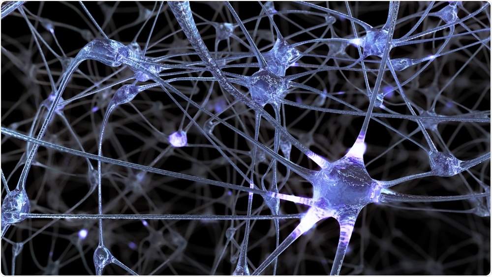
Neurons. Image credit: Vitaly Sosnovskiy / Shutterstock.com
How do sensory neurons work to detect friction and other tactile movements?
The glabrous skin of the human hand contains sensory neurons that change rapidly and slowly. They differ in the way they respond when stimulated. If you pick up a cup of coffee, the rapidly changing neurons at your fingertips will respond the very moment you grab it and then stop until you put the cup back on the table to which they set fire. Action again; but the slowly changing neurons will continue to fire electrical impulses as you contact the finger with the cup.
The rapidly and slowly changing neurons comprise four types of end organs: Meissner corpascles, Merkel cells, Pacinian and Ruffini corpuscles activated by various mechanical stimuli. The characteristics of neurons along with the positioning and anatomy of their end organs determine what type of stimulus executes best and whether a short-term or permanent response is aroused. With these four different types of mechanics, we can find small geometric details (e.g. edges, corners), delicate textures, vibrations, or layers of the skin.
We studied two of these four types of neurons, namely fast-changing type 1 neurons (FA-I) that make up Meissner’s corpuscles, and make slow-moving changes. type 1 (SA-I) which includes Merkel cells as these provide information on spatial detail. of objects and surfaces that our fingers touch.
Why has the sensitivity of one sensory neuron not been studied before?
Studying responses of a single tactile neuron in awake individuals requires a registration electrode to be inserted into a peripheral nerve. This method called microneurography is very challenging and only a few research organizations in the world use it and at the same time, there are so many scientific questions to be answered.
Despite the challenges of this approach, many studies on tactile information processing have been conducted over the years, and each ongoing outcome introduced new insights that will shape the next phase of research. The right time had to come for this investigation to take place. FA-I and SA-I neurons have infectious domains with several zones of maximum sensitivity (also referred to as “highly sensitive zones” or “subfields”) already described in 1978 by Roland Johansson (Quick sensation in human hand: infectious field characteristics of mechanical units in glabrous skin area. Johansson RS J Physiol (Lond). 1978) and then shown in one or two other studies over the years.
In 2014 Pruszynski and Johansson’s study (peripheral processing in first-order tactile neurons. Pruszynski JA, Johansson RS Nat Neurosci. 2014) showed for the first time that peripheral shape stimulus guidance regarding spatial arrangement of sub- areas of neurons affect neural response. Some marginal guides were preferred because they were well aligned with highly sensitive zones of neuron receptor domains. These results showed that the formation of subfields within a receptor field played a crucial role in the type of information presented and provided a new definition of the very high spatial acuity that humans exhibit when they handle objects.
That prompted the next step, which was to systematically study the subfields of the receptor field as well in the context of the spatial sensitivity of a single neuron. It has been known for a long time that the terminal organs of FA-I and SA-I neurons are located within papillary spines and we wanted to find out how much the neuronal responses are attached to the spines and how which were related to their spatial acuity of neurons.
Can you tell us how you did your most recent research on our consciousness?
We recorded electrical impulses from tactile neurons including the fingers of a dozen participants when a pattern of elevated dots examined their receptor domains. The dot pattern was wrapped around a rotating drum. There were 41 dots, spaced at least 7 mm apart, so that there was always only one dot crossing the receptor range; each dot had a specific position on the pattern (with 0.2 mm intervals) so that all the dots covered an 8 mm wide area on the skin that was perpendicular to the long axis of the finger. That allowed us to study the total infectious range of a neuron during one turn of the drum. Next, we created a sensitivity map of the receptor range of the neuron and analyzed its spatial sensitivity and strength of the neuron during different distances and directions of movement of the drum.
For some neurons the drum was rotated at three speeds: 15, 30, and 60 mm / s which is in the range of manual use in the world and for some neurons we also analyzed the effects of different directions scanning, mimicking back and forth movements. the finger.

Rubbing sensation. Image credit: Luma creative / Shutterstock.com
What did you find?
We found that the spatial sensitivity of highly sensitive zones fits around the width of a single papillary ridge (0.44 mm). That suggests that the subterranean end organs that measure the luminosity of individual ridges themselves suggest that papillary ridges are essential for discriminative rubbing. This changes how we think the tactile information is characterized by peripheral nerves.
We also show that the shape of highly sensitive zones is well preserved over time and over different speeds and motion directions. In other words the neural response is anchored to the ridges and no matter how many times we scan a surface, at what speed or in which direction, the spatial information we receive from the neurons in our fingerprints pretty much the same. .
How will your research help understand the body’s sensitivity to different stimuli?
Our findings offer an unambiguous explanation of why when handling objects with such high spatial clarity, it is something that cannot be explained by traditional, largely established models. on data from monkeys. There are ~ 210 receptor fields / cm2 of FA-I and SA-I neurons in our fingers and each has several subfields, spread over multiple ridges, which means that subfields belonging to many neurons are well between -woven.
Adding to that the fact that it is enough to avoid a single papillary ridge in order to elicit a response in one neuron allows us to think in detail how much information about the friction surface the brain receives.
It has been suggested that the surface texture information is carried in the vibrations that are aroused when our fingers slip over it.
However, it has been proven that our understanding of texture does not depend on how fast we move our hand over the surface, but the frequency of the vibrations will depend on that. So there needs to be additional equipment that allows us to feel the texture no matter how fast the movement. We show that the information in the FA-I and SA-I neurons was retained over different scan speeds indicating that the spatial information may contribute to texture understanding.
What are the next steps in your research?
To determine the sensitivity of the receptor domains of the neuron we have used one stimulus dot at a time. We know that there are several highly sensitive zones within a receptor range, so now we would like to know if any interaction between the cloud activity arises in different subfields if stimulated we do them at the same time (using more than one stimulus dot at a time).
We would like to examine the way in which the FA-I and SA-I neurons combine signals from their subfields.
About Dr. Ewa Jarocka
Dr. Jarocka works as a senior research engineer at the Department of Integrative Medical Biology at Umeå University.
Dr. Jarocka’s background is in physiotherapy but since coming to Sweden for post-doc, she has been involved in microneurographic studies of sensory information from both glabrous and hairy skin and muscle spindles – receptors located in our muscles.