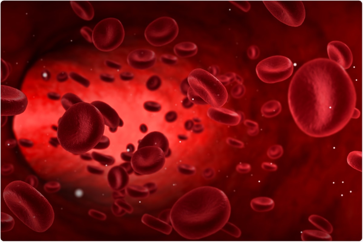
Red blood cells are anucleate cells, the most abundant cell type in the body, present in all substances, with a lifespan of 120 days. They also lack major class I histocompatibility complex molecules, and therefore, type O negative RBCs can be used with all patients. This automatically makes them ideal as delivery vehicles for therapeutic molecules, for a range of conditions, including coronavirus pandemic 2019 (COVID-19).
In a new study, released on the bioRxiv* preprint server, a team of researchers is studying the antiviral ability of red blood cells against severe acute respiratory coronavirus 2 (SARS-CoV-2) syndrome, the causative pathogen of COVID-19.
Achieving high levels of HIV-1 receptors
Engineering RBCs can be used to design viral traps, which attract viruses to attach and infect. This is achieved by enabling viral receptors to be displayed on their surface. The viruses that infect the RBCs cannot reproduce due to the lack of nucleic acid, which protects the host target cells from infection.
To express proteins, the cell must have translation mechanisms, which are absent within mature RBCs. To do this, erythroid progenitor cells must be engineered before differentiation. During the maturation process, transgene expression is usually inhibited by transcriptional disruption, mechanisms that control protein synthesis from transcribed genes, and protein breakdown not normally found in RBCs.
Transgene sentence
To achieve this, the researchers combined a transgenic sensing system combined with transgene codon optimization, to allow the RBCs to express HIV-1 CD4 and CCR5 receptors at high levels. This transformed the enucleated RBCs into viral traps that are powerful inhibitors of HIV-1 infection.
The researchers first applied an in vitro protocol to differentiate human hemopoietic CD34 + stem cells (HSCs) into reticulocytes (undifferentiated RBCs that still contain ribosomal RNA, so they can still between -translation of proteins, but lack of nuclei). HSC proliferation was followed by insertion of the ‘foreign’ CD4 or CCR5 genes into the erythroid progenitor cells by lentiviral vectors.
In addition, the researchers synthesized a gene to express fusion protein CD4-glycophorin A (CD4-GpA). This protein is composed of extracellular CD1 D1D2 domains attached to the N-terminal end of the common RBC protein, GpA. This was to allow single-domain antibodies to be expressed in RBCs. CD4 is a single-stranded, and CCR5 is a multimodal transmembrane protein. This protocol is only suitable for CD4, and not CCR5
They found that the use of the CMV promoter or ubiquitous promoters led to a low transgene feeling. So they moved to using a specific erythroid stimulant using the CCL-βAS3-FB lentivirus. This vector contains elements that upstream beta-globin expression during RBCn development. These vector elements include β-CD4, β-CD4-GpA, and β-CCR5.
The result was a significant increase in CD4 expression, a smaller increase in CCR5, and no change in CD4-GpA. This was due to the small number of ribosomal RNAs and intracellular RNAs, which restricted the expression of transgenes in the different RBCs.
Codon optimization
To address this, they made full use of the transgene codons, ensuring that they generated cDNA sequences that improved the sensitivity of all transgenes.
These engineered cells were effectively differentiated into enucleated RBCs, almost all of which expressed GpA, and one in three reported high levels of CD4 and CCR5 similar to the CD4 + T cells. approximately 6% of CD4-CCR5-RBCs are infected with the HIV-1 virus test, compared with 0.3% or less of control RBCs or CD4 + RBCs. Overall, therefore, this amounts to about one-fifth of CD4-CCR5-RBCs.
With CD4-CXCR4-RBCs, higher disease rates were observed. Infection levels were low with CD4-GpA-CCR5, or CD4-GpA-CXCR4 cells. Continue this even by adding the CD4 D3D4 fields. Perhaps the reason for such a low frequency of infection is the inability of CD4-GpA to co-localize with CCR5 and CXCR4 receptors, since GpA cannot localize to lipid subdomains, unlike CD4.
Engineering RBCs strongly neutralize HIV-1
The potential for viral capture was assessed by an HIV-1 neutralization assay. This showed that it is possible to achieve therapeutic concentration in vivo, since the semi-maximum barrier density (IC50) for HIV-1 pseudoviruses was 1.7×10⁶ RBCs / mL, which is about 0.03% of RBC concentration in human blood. The higher sensitivity level of CD4-GpA significantly increased neutralization activity.
Lower neutralization activity occurred when CCR5 was synthesized by CD4 or CD4-GpA RBC, prior to 2–3 hours, possibly due to CCR5 causing a small drop in the level of CD4-GpA and CD4 sensitivity. However, in vivo studies will be needed to demonstrate the potential for benefit by CCR5 co-administration of RBC viral arrests.
Their work on virus-like nanoparticles yielding concentrations of CD4 (CD4-VLP) showed that the ability of these particles to interact with HIV-1 envelope protein trimers allowed different types of HIV-1 neutralize them, while also preventing viral escape. in vivo. Interestingly, such interactions increase the potency of CD4-VLPs more than 10,000 times compared to soluble CD4 and CD4-Ig inhibitors. The present study shows that the same level of efficacy can be expected by the use of RBC and CD4-VLP viral receptors through high-avidity interactions with HIV-1 Env antigens.
Stable genera of RBC viral traps
This strategy was then used to produce erythroid progenitor lines that generate strong RBC viral arrests against HIV-1 and SARS-CoV-2. To ensure that these viral arrests were continuously induced, the researchers modified the immortalized erythroblast cell line (BEL-A) to express CD4-GpA at consistently high levels. They found that they effectively differentiated to enucleated RBCs in more than half of the cells, while continuing to express the engineered antigen. These cells potently strengthened HIV-1 infection with an IC50 of 2.1×10⁷ RBCan / mL.
SARS-CoV-2 uses the angiotensin converting cell enzyme 2 (ACE2) as its entry receptor. The extracellular range of ACE2 was bound to GpA to form a chimney protein, and a BEL-165 cell line was engineered to express this protein. When this was exposed to the lentivirus-based SARS-CoV-2 pseudovirus, the researchers found that the virus was highly neutralized, with an IC50 of 7×10⁵ RBCan / mL.
These findings suggest that the cell lines used to induce such viral traps from RBCs, expressing marked host receptors, can be rapidly generated. “RBC viral arrests have the potential to be powerful antiviral agents against a range of viruses.” They can persist in the body for 120 days, thus ensuring ongoing control of HIV-1 infection.
* Important message
bioRxiv publishes preliminary scientific reports that are not peer-reviewed and, therefore, should not be seen as final, guiding health-related clinical / behavioral practice, or treated as fixed information.