A look at RNA tells us what our genes want our cells to do, and scientists say that looking directly at the RNA of brain tumor cells seems to produce provide reasonable, effective evidence to better classify tumors and the most effective treatments.
Gliomas are the most common type of brain tumor in adults, they have a wide range of possible outcomes and three subtypes, from the more commonly treatable astrocytomas and oligodendrogliomas to the usually fatal glioblastomas.
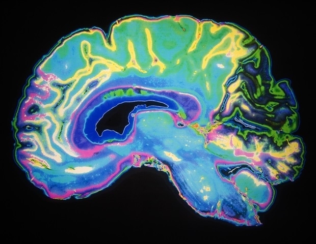
Georgia Medical College scientists report in the journal Scientific Reports that their approach, which produces an image called a tumor in particular, is capable of identifying some of the worst tumors, says Paul MH Tran, an MD / PhD student.
Gliomas are currently classified by histology, specifically the shape, or morphology, pathologists see when they look at the cancerous cells under a microscope, as well as identify mutations that are gene causing cancer.
“We are adding a third approach,” said Dr. Jin-Xiong She, director of the MCG Center for Biotechnology and Genomic Medicine, Eminent Alliance Scholar in Genomic Medicine and corresponding author of the study. Tran, who is doing his PhD work in She’s lab, is the first author.
While most patients have performed both conventional classification methods, results are sometimes inconsistent between the two groups, as traditional pathology detects glioblastoma cancer when the study did not perform a mutation and otherwise, and even when two pathologists look at the same brain tumor cells under a microscope, the scientists say.
To take a closer look at what a cancer cell is up to, they chose to look at untested gene expression, specifically the one-step downstream RNA, which shows where the cell is in the head. DNA expression is equivalent to RNA in that DNA produces RNA, which makes proteins, which determine cell function. One way cancer thrives is by altering gene expression, turning some up and others down or off.
They suspected that the new method would provide additional insight into the tumor, continue to evaluate the effectiveness of existing classification methods and are likely to identify new treatment targets.
RNA would be a detail of what is high and what is currently low in these glial cells as they are removed from the body. They are actually looking at how many replicas of relevant RNA genes are making. That gene expression usually determines everything from your hair color to how much weight you are measuring. The transcription profile counts the number of copies of each gene you have in the cell. “
Paul Tran, MD / PhD Student, Georgia College of Medicine at Augusta University
The glial genes, which are responsible for supporting neurons, have a tightly regulated gene expression that allows them to do so. With cancer, one of the first things that happens is how many RNA copies of each gene the cells undergo changes with which important cell function changes. “You change the expression of a gene to be something different,” she says.
Transcriptomic profiling begins like the other methods with a tumor sample from the surgeon, but then goes through an automated process to extract RNA, which is inserted into an instrument that reads gene expression levels for the different genes. The huge amount of data that is then generated is fed into a developed Tran machine learning algorithm, which makes up the most likely glioma subtype and associated prognosis.
They started with the Cancer Genome Atlas (TCGA) program and the Brain Molecular Neoplasia Database (REMBRANDT), two datasets that had already done the job of looking at RNA and also providing clinical information associated, including outcomes on more than 1,400 patients with gliomas. Tran, She and their colleagues used their algorithm to find gene expression patterns and used these patterns to classify all glioma patients without any other support. They then compared the three main glioma subtypes that emerged with standard classification methods.
Their transcription classification was about 90% in agreement with the traditional approach looking at cells under a microscope and about 93% agreeing with looking at genetic mutations, she says. They found about a 16% difference between the two standard measures.
“About 10-15% of patients disagree about the three methods,” she says, but notes that the most accurate study of the three should be because the their way better than the others at predicting survival.
And the inconsistencies they found between classification methods may be important for some patients despite close percentages.
“We found that our method may have some advantages because we found that some patients had a worse prognosis that could be identified by our method, but not by the other approaches, “Tran says.
For example, patients with astrocytoma or oligodendroglioma usually have a mutation in a gene called IDH, or isocitrate dehydrogenase, which is usually more suitable for treatment and has better survival rates than glioblastomas. But they also found that even some lower-grade gliomas with this IDH mutation can progress to something called secondary glioblastoma, something that is not available with the other two methods. IDH mutation is very rare in primary glioblastomas, Tran notes.
Using the conventional methods, which look at a small picture in time, those astrocytomas that progress to more lethal glioblastomas were classified as less severe tumors in 27 patients. “That wonder of progress is well known but we have a better way of highlighting those issues,” Tran says.
Further analysis also found that approximately 20% of patients with a worse prognosis had mutations in the TERT gene promotion category. The TERT gene is known for making telomerases, an enzyme that allows our chromosomes to stay healthy, while being known to decrease with age. The action of TERT is known to be overtaken by cancer to allow the endless cell proliferation that is a hallmark of cancer. This mutation is not usually present in glioma that begins as a more invasive glioblastoma, and means that a mutation in a TERT promoter is important in glioma progression, they say.
“The effect would be if we have protectors or something else that targets the TERT gene, you may be able to prevent some of these cases from having a worse prognosis,” Tran explains say.
These findings also highlight the strengths of the different classification methods, in this case suggesting that classification by mutation may not elevate these most aggressive brain tumors the new method of transcription, as well as the oldest method of looking at the cancer cells under a microscope. , better at making this important difference.
“It is known that a certain proportion of your low-grade gliomas can progress to glioblastoma and these are some of the ones that could be identified by the original techniques,” Tran says. Using our gene expression method, we found them even though some of them have IDH mutation. “
All of these changes with organizations like the World Health Organization calling for better ways for patients with IDH to determine a poor prognosis, they write. Other differences include some glioblastomas with the normal IDH gene carrying one of the worst prognoses for gliomas, but there is a subgroup of glioblastomas that function more like astrocytes and tend to carry a better prognosis.
Now that the MCG team has a better indication of which patients have a worse prognosis, the next steps include finding out why and perhaps what can be done.
In addition to prognosis accuracy, the second way to evaluate a tumor classification method is whether it points you toward better treatment options, she says, that they are now moving towards. It is noteworthy that most drugs and many of our activities, such as exercise and what we eat, alter RNA expression.
“Right now, if anyone gives us RNA sensory data from patients anywhere in the world, we can quickly tell them what the glioma subtype is most likely to be,” Tran says. As equipment that can monitor the sensitivity of RNA becomes more widespread, transcription accounts should become more widely available, they say.
Gliomas are tumors of glial cells – which include astrocytes, oligodendrocytes and microglial cells – brain cells that are larger than neurons and have a normal function of circulating and supporting neurons.
Identification of IDH gene mutations in the cells has already made normal glioma classification more systematic, the experts say. The mutation can be identified by staining the biopsy slide or by applying a layer for it.
Much progress has also been made in using machine learning to self-examine and refute the cancer and subtyping diagnoses they write about, including glioblastomas. Glioblastomas have been identified using transcription-based analysis but not all gliomas, as per the current study.
Like most genes, the IDH gene normally carries many functions in the body, involving the processing of glucose and other metabolites for several cell types. But when inverted, it can damage cells, triggering factors such as reactive oxygen species, which damage the DNA and contribute to cancer and other diseases. These mutations can occur with age and / or environmental exposures. IDH inhibitors are in clinical trials for a number of cancers including gliomas.
More insight is also emerging into the important DNA methylation that occurs in cancer, which alters gene expression, leading to changes such as suppression of tumor suppressor genes and the development of out genetic mutations that cause cancer.
Source:
Georgia Medical College at Augusta University