Our knowledge of Alzheimer’s disease has grown rapidly in the last few decades but it has been difficult to translate basic findings about the disease into new treatments. Researchers at the California National Center for Priority Research at the University of California, Davis have now developed a model of the early stages of Alzheimer’s disease in rhesus macaques.
The macaque model, published on March 18 in the magazine Alzheimer’s & Dementia: Journal of the Alzheimer’s Society could better explore new therapies.
The model was developed by the laboratory of Professor John Morrison of the CNPRC, in collaboration with Professor Jeffrey Kordower of Rush University Medical Center and Paramita Chakrabarty, an assistant professor at the University of Florida.
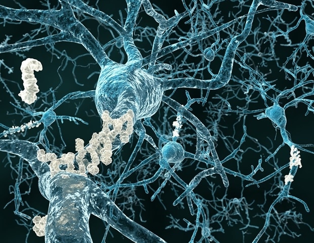
Alzheimer’s disease is thought to be caused by a misfolding of the tau and amyloid proteins. Uncomplicated proteins spread through the brain, leading to inflammation and cell death. Tau proteins are commonly found in neurons of the brain and central nervous system, but not elsewhere.
Researchers believe that decades may pass between the silent onset of the disease and the first signs of mental decline. Understanding what happens over the years may be fundamental in preventing or reversing the symptoms of Alzheimer’s disease.
But it is difficult to study therapeutic strategies without a powerful animal model that mimics the human condition as closely as possible, Morrison said. Much research has focused on transgenic mice expressing a human version of amyloid or tau proteins, but these studies have found it difficult to translate into new therapies.
New translation modules required
Humans and monkeys have two types of tau protein in their brains, but rodents have only one, said Danielle Beckman, a CNPRC postdoctoral researcher and first author of the paper.
“We think the macaque is a better model, because it exerts the same versions of tau in the brain as humans,” she said.
Mice also lack specific areas of the neocortex such as the prefrontal cortex, an area of the human brain that is highly vulnerable to Alzheimer’s disease. Pre-horizontal cortex is present in rhesus macaques and is extremely important for mental functions both in humans and in monkeys. There is an urgent need for new and improved animal models for Alzheimer’s disease that can stand between mouse models and human clinical trials, Beckman said.
Chakrabarty and his colleagues created strains of the human tau gene with mutations that cause mutation, wrapped in viral material. These vectors have been introduced into the macas rhesus, in a brain area called the entorhinal cortex, which is highly vulnerable in Alzheimer’s disease.
Within three months, they could see that undigested tau proteins had spread to other parts of the animal’s brain. They found a mismatch of both introduced human tau mutant proteins and of the tau proteins themselves.
The diffusion pattern showed that tau-based pathology followed precise connections of the entorhinal cortex and that pathological tau seeding could pass from one area to the next through synaptic connections. This ability to spread through brain circuits leads to damage to cortical areas that are dependent on higher level experience far from the entorhinal cortex, “
John Morrison, Professor, CNPRC
The same team has previously established the release of complex amyloid proteins in macaques, representing very early stages of Alzheimer’s disease, by injecting short pieces of defective amyloid. The similarly new tau protein model represents a moderate stage of the disease, Beckman said.
“We think this represents a reduced rate, but before cell death occurs far and wide,” she said.
The next plan researchers will test whether behavioral changes similar to human Alzheimer’s disease develop in the rhesus macaque model. If so, it could be used to test for prevention of inflammation or inflammation.
“We’ve been working to develop these models for the last four years,” said Morrison, “I don’t think you could have done this without a great collaborative team and the extensive facilities of the National Primate Research Center. “
Source:
University of California, Davis